Welcome everybody,today we would like to explain about Protein Data Bank.
The Protein Bank (PDB) is a repository for the three-dimensional structural data for large biological molecules, such as proteins and nucleic acids. The data, typically obtained by X-ray crystallography or NMR spectroscopy and submitted by biologists and biochemists from around the world.
X-Ray Crystallography
- A target is selected for structure determination
- The material is purified and then crystallized
- Crystals are put in front of an X-ray beam and diffracted intensities are collected on a detector
- These data are then analyzed using different computational methods

Data Deposition
PDB Extract Tool
- pdb_extract can extract information from the output of standard crystallographic programs.
- It merges the information into mmCIF files at each step of the structure-determination process.
- These mmCIF are then ready for validation and deposition.
AutoDep Input Tool
- ADIT is used for assembling, editing, validating and deposition structural data.
- It is built on top of the mmCIF dictionary.
PDB Validation Suite
- Creates reports based upon the validation results.
- Also calculates derived information that could be used for assessing the quality of a structure.
For more information on process of data deposition,click here.
Examples of protein structure images with their description:
| Classification | Alpha amylase |
| Structure weight | 48016.77 |
| Molecule | ALPHA-1,4-GLUCAN-4-GLUCANOHYDROLASE |
| Polymer | 1 |
| Type | Protein |
| Length | 425 |
| Chains | A |
| Organism | Bacillus subtilis |
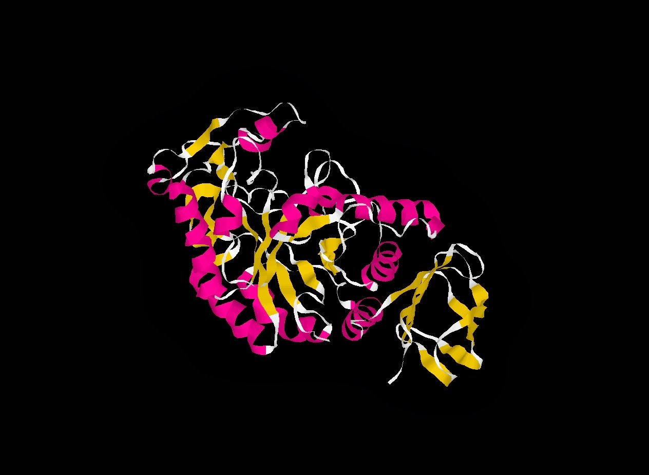
THE GEOMETRY OF THE REACTIVE SITE AND OF THE PEPTIDE GROUPS IN TRYPSIN, TRYPSINOGEN AND ITS COMPLEXES WITH INHIBITORS
| Classification | Complex (proteinase/inhibitor)) |
| Structure weight | 29892.08 |
| Molecule | BETA-TRYPSIN |
| Polymer | 1 |
| Type | Protein |
| Length | 223 |
| Chains | E |
| Organism | Bos taurus |
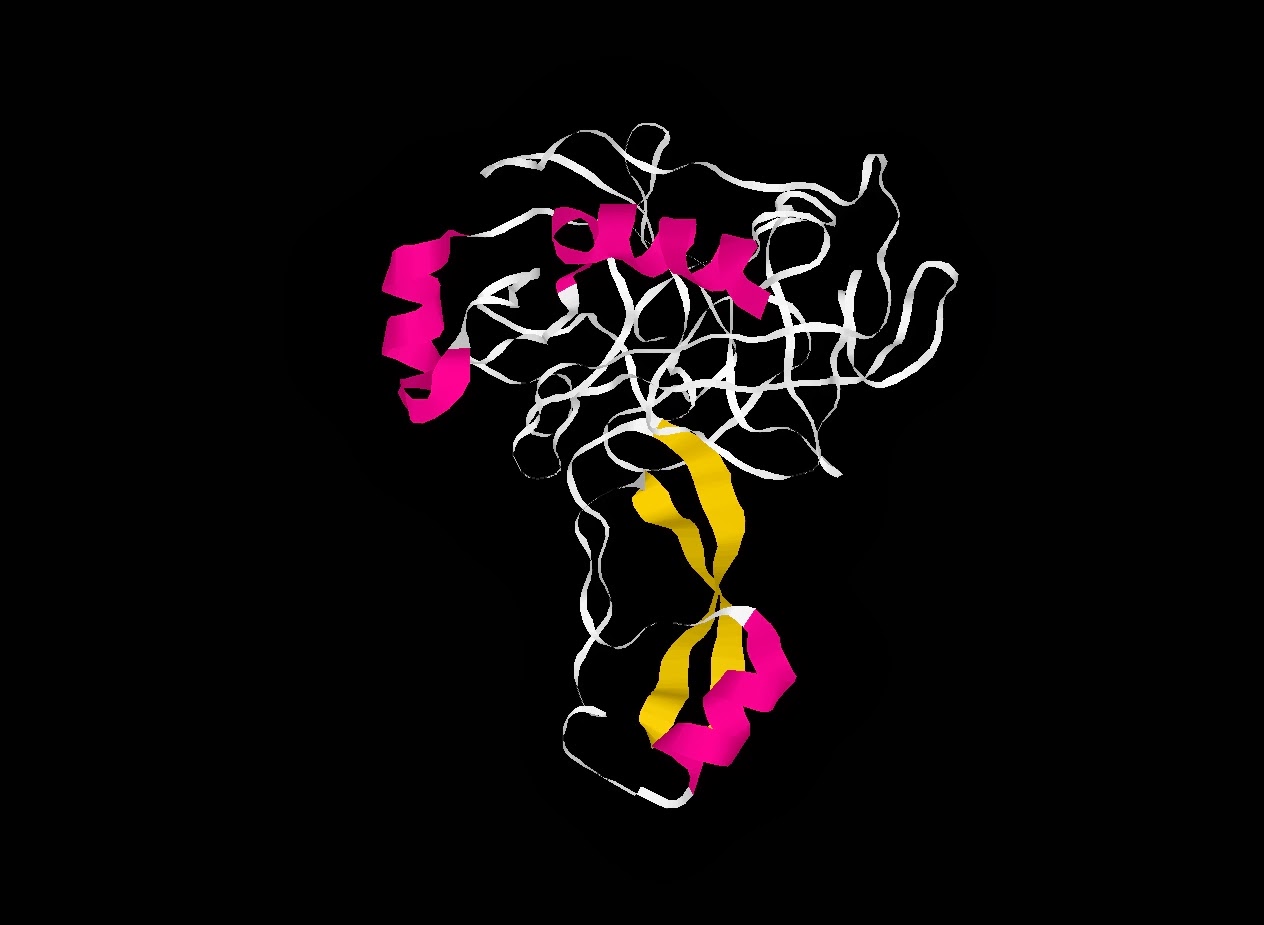
X-RAY ANALYSES OF ASPARTIC PROTEASES. II. THREE-DIMENSIONAL STRUCTURE OF THE HEXAGONAL CRYSTAL FORM OF PORCINE PEPSIN AT 2.3 ANGSTROMS RESOLUTION

| Classification | Hydrolase(acid Proteinase) |
| Structure weight | 34469.80 |
| Molecule | PEPSIN |
| Polymer | 1 |
| Type | Protein |
| Length | 326 |
| Chains | A |
| Organism | Sus scrofa |
SOLUTION STRUCTURE OF HTRA PDZ DOMAIN FROM STREPTOCOCCUS PNEUMONIA
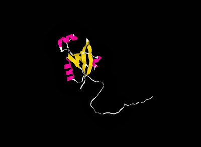

| Classification | Protein Binding |
| Structure weight | 14890.90 |
| Molecule | Putative serine protease |
| Polymer | 1 |
| Type | Protein |
| Length | 134 |
| Chains | A |
| Organism | Streptococcus pneumoniae |
CRYSTAL STRUCTURE OF BOVINE PANCREATIC CARBOXYPEPTIDASE A COMPLEXED WITH AMINOCARBONYLPHENYLALANINE AT 1.75 A

| Classification | Carboxypeptidase |
| Structure weight | 138865.59 |
| Molecule | CARBOXYPEPTIDASE A |
| Polymer | 1 |
| Type | Protein |
| Length | 307 |
| Chains | A, B, D, E |
| Organism | Bos taurus |